RELIABLE
RESULTS
DIAGNOSTICS
Where further investigation is identified to help assess your injury, we use a selection of leading diagnostic services from the best nationwide providers. See the below guides for the diagnostic services we provide.
-
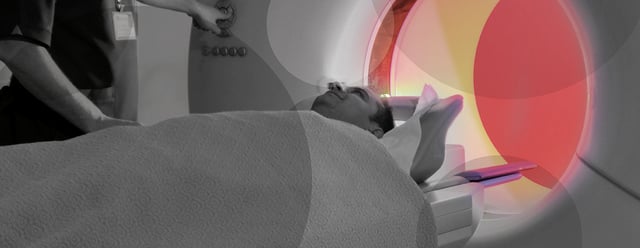
MAGNETIC RESONANCE IMAGING (MRI)
WHAT IS AN MRI?
MRI is a type of scan that uses strong magnetic waves to produce 3D images of muscles, ligaments, joints or other parts of the human body, so that a more accurate diagnosis can be reached and the right treatment plan can be developed.
Unlike an X-Ray, there’s no exposure to radiation from an MRI scan, and apart from a bit of noise during the scan, and the feeling of being inside a small tunnel, it usually causes no discomfort for patients.
An MRI scan is often a vital part of diagnosing a variety of conditions, some of which may have obvious symptoms, whilst some may not.
Typical conditions identified by MRI scans include;- Joint and muscle pain
- Torn ligaments
- Spinal cord and intervertebral disc injury
When would you need it?
You may have neurological symptoms in your legs i.e. pins and needles, numbness following a back injury that isn’t responding to physiotherapy. MRI scanning can ascertain the extent of nerve related pathology in order that the most appropriate treatment can begin as soon as possible.
How it works
For some conditions, a coloured dye may be injected into the patient, so that the MRI scanner can build a more detailed picture, showing liquid moving in the body. This is called an MRI Arthrogram, and is particularly good in determining tears and other lesions.
-
ARTHOSCOPY (A TYPE OF KEYHOLE SURGERY)
WHAT IS ARTHROSCOPY?
Arthroscopy is a type of keyhole surgery, often used to diagnose and treat problems with joints, especially in the knees, ankles, shoulders, elbows, wrist and hips. It is a relatively simple process and can be extremely useful in identifying the underlying cause of persistent joint pain, swelling or stiffness when scans haven’t been able to identify the cause.
Once a local anaesthetic has been applied, a small camera can be inserted and data is gathered on the potential problem. Sometimes a small amount of liquid may be inserted so that the specialist can see the joint in an expanded view, as this can help better treat any problems.
Arthroscopy is particularly useful in treating a range of joint problems and conditions, including:
- Removing fragments of loose bone or cartilage
- Draining away any excess fluid
- Repairing cartilage damage
- Treating arthritis, frozen shoulder or carpal tunnel syndrome
When would you need it?
You might be suffering knee or hip pain. Torn ligaments are another injury where keyhole investigation is frequently used.
If you are suffering ongoing pain in one of your joints then Arthroscopy offers a chance to physically look inside the joint in detail and record the images on camera. A specialist can then assess the condition and provide the correct treatment.
How it works
This procedure can work well for those who have suffered a trauma type of injury. Once assessed, a patient would attend a clinic and have the area affected prepped for arthroscopy.
Depending on the nature of the problem, and its location on the patient’s body, the examination via camera could take between 30 to 90 minutes.
-
BONE SCANNING
WHAT IS BONE SCANNING?
A bone scan is a type of imaging test, similar to a Computed Tomography (CT) or X-ray in some ways. A small tracer dye is put into the bloodstream, which works its way around the body. This radioactive dye helps to identify how effectively a broken bone is healing by looking at the metabolism of the bone structure. In other words, how the bone is rebuilding and knitting itself back together.
The tracer dye is absorbed more rapidly by bone which is repairing itself, so potential fractures would show up as `hot spots’ on the imaging process. This type of scan can also be extremely useful in spotting fractures, or bone problems that might have been missed during routine X-rays.
Bone scans are especially useful for identifying problems such as;
- Lower back pain and bone abnormalities
- Fractures which are not healing well
- Small problems within complex bone structures, such as the spine or foot
- Arthritis
- Infection involving the bone
When would you need it?
Bone scans are often beneficial in identifying stress fractures. A bone scan could also help diagnose the root cause of ongoing back or joint pain, and help identify abnormal bone growth or a bone which is not healing properly.
How it works
Typical treatment would be the Three Phase Bone Scan. A tracer dye is injected to build up images of the affected area. Any radioactive material from the tracer test is naturally removed from the body within two days. The procedure takes about one hour and you will have to remove any jewellery.
-
COMPUTED TOMOGRAHPY (CT)
WHAT IS A CT SCAN?
A CT scan is essentially an X-Ray in 3D. The scanner rotates around the patient, building up a series of overlapping images, which give a better view of any potential problems or injuries. Sometimes known as a CAT scan, these images can aid rapid diagnosis of a variety of conditions by looking at the density and structure of tissue inside the body.
Unlike an MRI scanner, where you are placed within a tunnel, a CT scanner consists of a doughnut shaped machine and therefore you should not feel claustrophobic.
Pregnant women should avoid having a CT scan, but for most adults, it is perfectly safe as the benefits strongly outweigh any risks.
A CT scan can be useful in the following cases;
- Broken bones
- Damage to internal organs
- Examination of the chest, abdomen and pelvis
- Damage to the spine and injuries to the hands, feet and other skeletal structures
When would you need it?
If you have suffered injuries in a road accident, suffered a fall, or perhaps sustained a blow to the head, a CT scan can help diagnose problems quickly and effectively. The scan itself is painless, and you should wear loose clothing with no zips. Jewellery should also be removed.
How is it carried out?
The scan usually takes approximately 10 to 30 minutes, dependent on the part of the body that is being scanned, the number of pictures taken and the different angles required.
-
ELECTROCARDIOGRAM (ECG)
WHAT IS AN ECG?
An ECG is a long-established method of diagnosing heart problems and has been standard practice for many years now. The test is fairly quick and usually consists of 12 wires being attached to the patient. These are mainly to the chest area, but also include the arms and legs so that a comprehensive analysis of heart performance and blood flow can be recorded.
Typical problems that might require an ECG would include:
- Heart or chest injuries following a road accident
- Chest pains
- Dizziness or fainting
When would you need it?
A 12 point ECG is one of the most effective ways to diagnose a potentially serious chest injury following a road accident. In some cases, further testing may be required within 48 hours of the initial ECG.
An accident can place great stress on the heart, even if the injuries seem initially superficial, so it is important to assess how the heart performs when resting and when the person is active.
How it works
An ECG is usually completed at hospital but can be taken at GP Surgeries or Clinics. It only takes minutes to attach the wires and then the heartbeat can be monitored. Usually, the patients `resting’ heart-rate tested first whilst they’re lying down.
A more involved ECG, often called a Treadmill Test, might take around 30 minutes to complete and this involves increasing the heart-rate through mild exercise. The patient is required to walk on a treadmill whilst slowly gather speed and sometimes features a slight incline.
-
NERVE CONDUCTION STUDY (NCS)
WHAT IS NCS?
Nerve conduction studies help to test how well and how fast nerves conduct electrical signals.
Nerve damage can be caused by things like road accidents, falling from height or being hit by an object. An NCS is a way of seeing how well nerves are transmitting electrical impulses around the body. The simple procedure attaches two electrodes to the skin where the nerve damage has occurred. An impulse is sent between the terminals to test if the nerve is working properly. Then the signal speed between the two electrodes is calculated. It’s very similar to a speed camera.
An NCS test is ideal for diagnosing problems such as;
- Trapped nerves i.e. carpal tunnel syndrome
- To assess nerve damage following an injury
- Test for conditions affecting the nervous system
- Check for damage to nerves caused by diabetes
When would you need it?
NCS is able to detect problems with the optic nerve, or peripheral nerve damage (tingling or numbness in fingers) in some patients.
If you suffer any kind of numb feeling, especially following an accident, then an NCS test is a good way to investigate.
How it works
A typical NCS test can take between 30 minutes and 90 minutes depending on the complexity problem. In most cases, the initial electrode test is followed up immediately with a fine needle test, involving a needle being inserted into the patient, testing nerve conduction.
The NCS tests would normally take place at a local hospital although there are some clinics who deliver these examinations using hand-held devices.
-
SPECT SCAN
WHAT IS A SPECT SCAN?
A SPECT scan stands for Single Photon Emission Computed Tomography, and essential it is an enhanced imaging technique, that is made up of 2 separate components, a SPECT scan and a CT scan, whereby the images from each scan are fused together to provide more accurate information in identifying and localising a problem.
A small amount of radioactive tracer solution is injected into the patient to determine how an area of the body is functioning. As the tracer works through the brain, or other parts of the body, a series of images can be taken using three cameras rotating around the patient.
SPECT analysis is used for diagnosing problems such as;
- Head injury following an accident
- Spinal pain
- Intervertebral disc, facet joint and sacro-iliac joint inflammation
- Hidden bone fractures
- Cognitive decline or memory disorder
- Follow up testing after post-accident treatment
When would you need it?
Many hospitals find that SPECT analysis is a useful complementary procedure, especially when used alongside Magnetic Resonance Imagining (MRI) or CT scanning. It often helps to provide a complete picture when trying to assess ongoing pain, or niggling injuries.
The aftermath of an accident can affect the brain in a variety of ways, so SPECT scanning looks deeper into the causes behind mood swings, depression or similar post-traumatic conditions.
How it works
SPECT testing is relatively quick, taking around 20-30 minutes to complete. It is useful in two key areas; the diagnosis of head injury and as a follow-up examination after treatment for a variety of injuries. A SPECT test would usually take place at a local hospital as the machinery is fairly large. Patients should wear loose clothing and no jewellery.
-
ULTRASOUND
WHAT IS ULTRASOUND?
Ultrasound scanning is an accurate and cost-effective way of measuring bone structure, muscle tissue and identifying things like torn ligaments.
Unlike X-rays, there’s no radiation involved and many ultrasound scanners are far more portable than most medical machinery, so a scan can be performed in a variety of locations.
An ultrasound is particularly good at detecting slight changes in bone structure, especially when assessing tiny fractures or dislocations, which may have been missed during an initial examination.
An ultrasound would be useful in diagnosing the following problems;
- Sports related injuries like sprains and knee ligament problems
- Injuries sustained during a trip or fall from height
- Trapped nerves such as carpal tunnel syndrome
- Foreign bodies in soft tissues such as glass or splinters
When would you need it?
An ultrasound can be very effective in spotting scaphoid bone fractures in the wrist, which are typically suffered when someone falls or has a bicycle accident, for example. It’s also useful for detecting fractured ribs.
If you have an accident and receive treatment but find that you’re still suffering from the same pain then an ultrasound examination may help.
How it works
An ultrasound exam is carried out by a sonographer and can be completed at a clinic, or GP Surgery, rather than a hospital. It’s a pain-free process and takes just a few minutes in most cases.
A warm gel is applied and the operator then uses a transducer to form a 3D image of the affected area. If there’s a condition like a painful tendon or muscle, then the ultrasound examination can reveal how those tissues are behaving under load or rotation. This makes it an extremely useful diagnostic tool.
-
X-RAY
WHAT IS AN X-RAY?
An X-ray is a common imaging test that is familiar to most people and has been around for well over 100 years. In that time the radiation from a typical X-ray has decreased by a huge margin, making the process much safer. More importantly, image quality has also improved, making this a default diagnostic tool when assessing a range of injuries, not just broken bones.
An x-ray would be useful for identifying the following;
- Fractures
- Arthritis
- Chest pains or damage to internal organs
- Post-accident head injury
When would you need it?
- To examine an area where you’re experiencing pain or discomfort
- Monitor the progression of a diagnosed disease, such as osteoporosis
- Check how well a prescribed treatment is working
How it works
An X-ray is usually completed at a hospital for safety reasons, but some can be performed at GP surgeries or clinics.
It only takes minutes to complete and patients should wear loose clothing and no jewellery. Sometimes contrast liquids are used to enhance the images, often a barium drink which offers a better view as its travels around your body.
X-rays can be performed standing up or lying down on the table. Results will usually be available within a few days.